Ureteroscopy with Laser Lithotripsy
Ureteroscopy with Laser Lithotripsy is a minimally invasive procedure used to remove kidney stones located in the ureter or kidney. It involves the use of a small, flexible ureteroscope with a laser fiber that is passed through the urethra and bladder and into the ureter to break up the stone. The laser is used to fragment the stone into small pieces that can be easily passed out of the body through the urine.
Ureteroscopy with laser lithotripsy is typically performed as an outpatient procedure under local or general anesthesia. It is a safe and effective treatment option for kidney stones that are too large to pass on their own or cause significant pain and discomfort. The procedure has a high success rate and is associated with minimal complications.
After the procedure, patients may experience some mild discomfort or pain and may need pain medication. They may also need to drink plenty of fluids to help flush out any remaining stone fragments. Most patients are able to resume their normal activities within a few days.
Overall, ureteroscopy with laser lithotripsy is a highly effective and minimally invasive treatment option for kidney stones. It is a safe procedure that offers a quick recovery time and allows patients to return to their normal activities soon after the procedure. If you are suffering from kidney stones, consult with your urologist to determine if ureteroscopy with laser lithotripsy is the right treatment option for you.
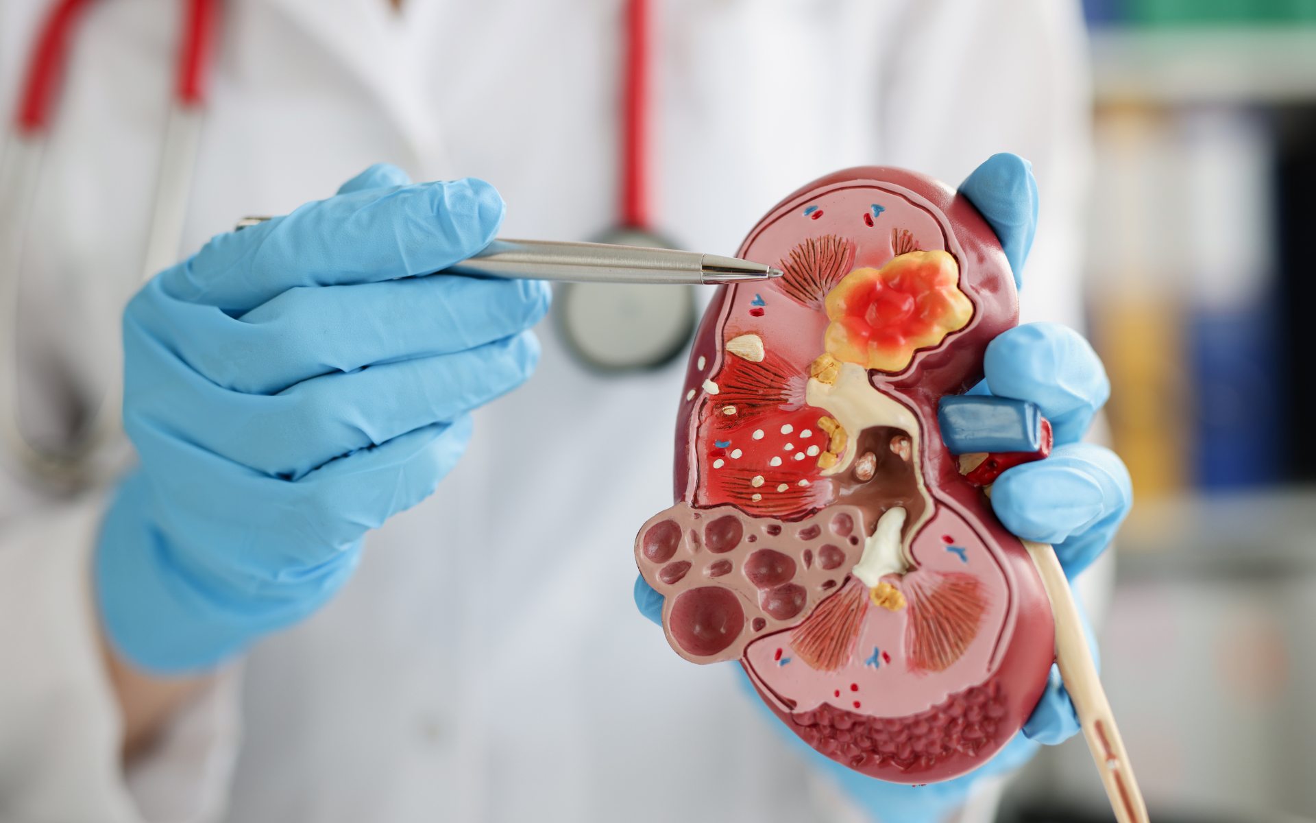
What is a Kidney

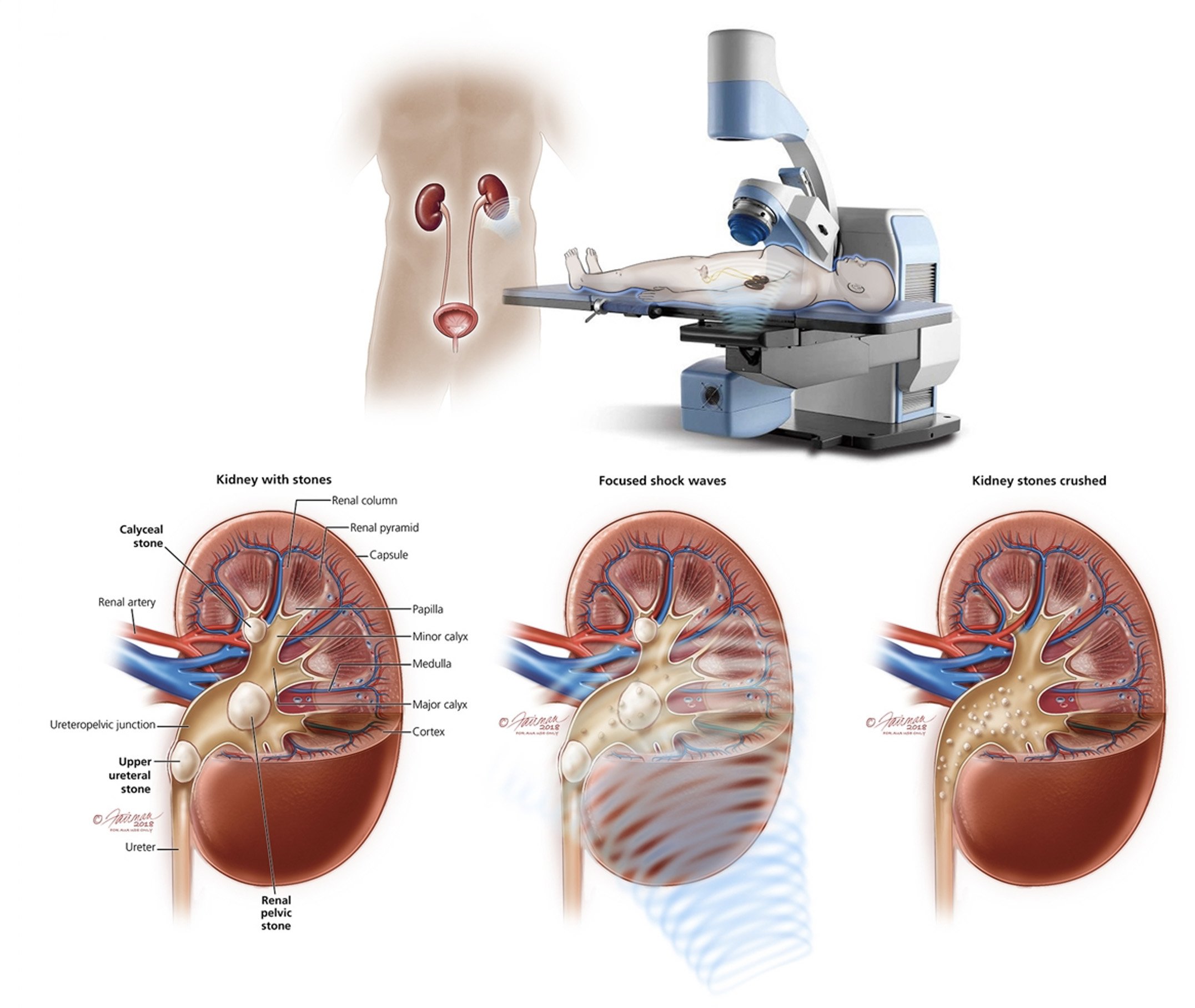
Each kidney is located on either side of the spine in the lower back, protected by the ribs. The outer layer of the kidney is called the cortex, while the inner layer is called the medulla. The kidney receives blood from the renal artery, which branches off from the aorta, and returns blood to the body through the renal vein.
The kidneys play a vital role in maintaining the overall health and well-being of the body. Any kidney damage or dysfunction can lead to a range of health issues, including fluid and electrolyte imbalances, hypertension, and kidney disease. Therefore, it is essential to maintain healthy kidney function, such as staying hydrated, maintaining a healthy diet, and avoiding smoking and excessive alcohol consumption.
What is Stone Disease
Stone disease, also known as urolithiasis or nephrolithiasis, is a condition where stones, also known as calculi, are formed in the urinary tract. These stones can develop anywhere in the urinary tract, including the kidneys, bladder, and ureters. The stones can be made up of different substances, including calcium, oxalate, uric acid, and cystine.
Kidney stones can be excruciating and can cause symptoms such as flank pain, blood in urine, nausea, vomiting and in some cases even sepsis. The size and location of the stone can also affect the symptoms experienced. Smaller stones can sometimes pass on their own, while larger stones may require medical intervention.
Several risk factors are associated with stone disease, including dehydration, high salt intake, and a diet high in oxalate-rich foods. Certain medical conditions such as gout, urinary tract infections, and certain genetic disorders can also increase the risk of developing stones. Treatment options for stone disease can vary depending on the size and location of the stone, as well as the individual’s overall health.
What is a Kidney Stone
A kidney stone is a hard, crystalline deposit inside the kidney or urinary tract. It comprises minerals and salts, and can vary in size and shape. Kidney stones can cause severe pain and discomfort when they pass through the urinary tract. If left untreated, they can also lead to complications such as urinary tract infections and kidney damage.
Kidney stones form when there are too many substances in the urine, such as calcium, oxalate, and uric acid. When these substances become concentrated in the urine, they can form crystals that stick together and grow into solid stone. Dehydration, certain medical conditions, and some medications can increase the risk of developing kidney stones.
Symptoms of kidney stones include severe pain in the back, side, or lower abdomen, nausea and vomiting, and pain or burning during urination. Small kidney stones may pass through the urinary tract independently, but larger stones may require medical treatment. Treatment options may include medications to relieve pain and help the stone pass, or surgical procedures to remove the stone or break it up into smaller pieces.
Types of Kidney Stones
There are several types of kidney stones, including:
Calcium stones
Calcium stones are the most common type of kidney stones, accounting for approximately 80% of all cases. They are usually composed of calcium oxalate or calcium phosphate. Calcium oxalate stones are more common than calcium phosphate stones and form when there is an excess of oxalate in the urine.
Oxalate is a naturally occurring substance found in many foods, including fruits, vegetables, and nuts. When there is too much oxalate in the urine, it can bind with calcium to form crystals that can eventually become stones. Calcium phosphate stones are less common and usually occur in people with a metabolic disorder or renal tubular acidosis.
Risk factors for calcium stones include a diet high in oxalate or salt, dehydration, family history, obesity, and certain medical conditions, such as hyperparathyroidism, inflammatory bowel disease, and urinary tract infections. Symptoms of calcium stones may include pain in the back, side, or abdomen, painful urination, and blood in the urine. Some people may experience no symptoms, and the stones may only be discovered during routine imaging tests.
Struvite stones

Symptoms of struvite stones are similar to those of other kidney stones, including severe pain in the back, side, or abdomen, nausea and vomiting, and difficulty passing urine. Diagnosis is made through imaging tests such as X-rays, CT scans, or ultrasounds. Treatment options for struvite stones include extracorporeal shockwave lithotripsy (ESWL), percutaneous nephrolithotomy (PCNL), and ureteroscopy with laser lithotripsy. Antibiotics are also prescribed to treat any underlying infections.
Uric acid stones
Uric acid stones are one of the most common kidney stones, accounting for approximately 10% of all kidney stones. These stones form when there is an excess of uric acid in the urine, which can happen due to various factors such as diet, genetics, and medical conditions like gout or chemotherapy.
Uric acid stones can be particularly painful and may require medical intervention to pass. Symptoms of uric acid stones can include intense pain in the back or side, nausea, vomiting, and blood in the urine. To diagnose uric acid stones, a healthcare provider may order imaging tests such as X-rays or CT scans, as well as urine and blood tests.
Cystine stones
Cystine stones are a type of kidney stone composed of cystine, an amino acid found naturally in the body. These stones form when there is an excess of cystine in the urine, forming crystals that can accumulate and grow over time.
Cystine stones are relatively rare, accounting for less than 1% of all kidney stones, but they can cause significant pain and discomfort for those who develop them. Because cystine is less soluble than other substances in the urine, it is more likely to form crystals and stones when urine becomes concentrated. People with a genetic condition known as cystinuria are at increased risk of developing cystine stones.
Mixed stones
Mixed stones, as the name suggests, are a combination of different kidney stones, typically calcium oxalate and uric acid stones. These stones can vary in size, shape, and composition, making them difficult to diagnose and treat.
Mixed stones can be particularly problematic because they may require a combination of treatments depending on their composition. In some cases, surgical intervention may be necessary to remove the stones. Diagnosing and managing mixed stones are important for preventing complications such as kidney damage or infection.Determining the type of kidney stone is important to develop an effective treatment plan.
Signs and Symptoms of Kidney Stones
Here are some signs and symptoms of kidney stones:
- Sharp pain in the back, side, or lower abdomen
- Painful urination
- Cloudy or foul-smelling urine
- Blood in the urine
- Nausea and vomiting
- Difficulty urinating or frequent urination
- Fever and chills (if an infection is present)
- Pain that comes and goes in waves and varies in intensity
- Pain that worsens when you move or change positions
- Pain that may spread to the groin or genital area
- Pink, red, or brown urine
- Feeling the need to urinate urgently or frequently.
Steps for a Ureteroscopy with Laser Lithotripsy
Below is a list of the steps for a Ureteroscopy with Laser Lithotripsy procedure:
-
- Preparation: The patient will be given anesthesia and positioned on the exam table with their legs in stirrups. The doctor will insert a ureteroscope, a long thin tube with a camera and light on the end, through the urethra and bladder to access the ureter and kidney.
- Locate the Stone: The doctor will use the camera on the ureteroscope to locate the kidney stone. Once the stone is found, the doctor will pass a small instrument through the ureteroscope to grab or break the stone.
- Laser Lithotripsy: If the stone is too large to remove the whole, the doctor will use a laser to break it into smaller pieces. The laser generates shock waves that break up the stone without harming the surrounding tissue. The fragments will be small enough to be removed with a basket or suction.
- Removal: The doctor will remove all the stone fragments from the kidney and ureter. If the stone is too large to remove completely during one procedure, the doctor may need to perform additional lithotripsy sessions.
- Stent: often after removal of stone from the kidney or ureter, a temporary stent will be placed to allow healing of the ureter and adequate drainage of urine from the kidney. This stent is usually removed in the office a few days after the procedure.
- Recovery: After the procedure, the patient will be taken to a recovery room and monitored for several hours before being discharged. Patients may experience discomfort and may be given pain medication to manage any pain. It is important to follow the doctor’s instructions on post-operative care, such as drinking plenty of fluids and avoiding strenuous activity, to promote healing and prevent complications.
- Follow-up: The patient will need to follow up with the doctor to ensure that all the stone fragments have been removed and to monitor for any potential complications. The doctor may recommend imaging tests, such as X-rays or ultrasounds, to confirm that the kidney stone has been completely removed.
Recovery From a Procedure
Recovery after a ureteroscopy laser lithotripsy depends on the size and location of the stone, as well as the overall health of the patient. In general, most patients can go home the same day or the day after the procedure. There may be some mild discomfort, including pain or burning sensations during urination, as well as some blood in the urine. Patients are typically advised to drink plenty of fluids to help flush out any remaining stone fragments and prevent infection.

